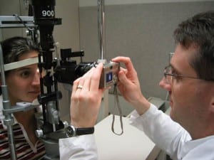
Slit Lamps are ophthalmic equipment that focus beams of light onto patients’ eyes. A bio-microscope is also required along with it. They are employed for the examination of anterior and posterior segments of humans’ eyes. Conjunctiva, cornea, eyelid, iris, natural crystalline and sclera are the various parts of an eye that can be obsereved with a Slit Lamp. A Slit Lamp magnifies the image of an eye which helps ophthalmologists in keen observation of the same and diagnosis of ill conditions of eyes. Utilities of a Slit Lamp has steadily increased due to invention of numerous accessories and add-ons for the device.
The device examines the eye of a patient in sitting position. A stable platform is required to support the chin of the patient so that beam from a Slit Lamp can be focussed properly. Direct focus of beam onto the eye is most commonly applied method of ophthalmologists. However, fluorescent-stained paper strip is also, at times used, for examination. Another examination procedure is named Direct Diffuse Illumination. This is mostly aopted when it is tough to get a clear images of the optical section. Survey of anterior segments, crystalline lens, surface of cornea, etc. are is done through this method.
Another procedure in which this specific ophthalmic equipment efficiently does Indirect Illumination. It is also employed for the examination of anterior chambers of corneas. Corneal scars, deposits, epithelia, infiltrates, stromal defects, etc. are examined through this method. However, in some cases, the method of illumination of optical section does not yield desired results. Examination of areas behind the crystalline lens is one of such situations. Retro Illumination is beneficial in these and similar situations. This is why almost all Slit Lamps have the ability to decentre the slit laterally.
Scattering Sclero-Corneal Illumination is a method which focuses wide beam focus onto limbal region of eyes at very low angle. Illumination or shadowing helps in the detection of scars, foreign bodies, inclusions and opacities. Fundus is observed with a Fundus camera. The refractive nature of the ocular media makes the observation of Fundus through a Slit Lamp impossible. However, if convex optics and concave optics are used together then this issue can be resolved. Concave lens produces upright intermediate virtual image of Fundus. Convex lenses produce inverted real images of objects in front. Thus, Slit Lamps are loaded with all that is required by ophthalmologists to treat eyes conditions.